GIVE NOW before 2025 ends—your gift will be doubled to help children in need. Click here to 2x your impact!

Ranked nationally in pediatric care.
Arkansas Children's provides right-sized care for your child. U.S. News & World Report has ranked Arkansas Children's in seven specialties for 2025-2026.

It's easier than ever to sign up for MyChart.
Sign up online to quickly and easily manage your child's medical information and connect with us whenever you need.
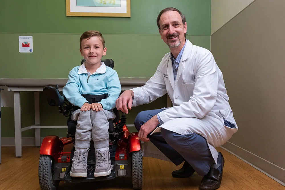
We're focused on improving child health through exceptional patient care, groundbreaking research, continuing education, and outreach and prevention.

When it comes to your child, every emergency is a big deal.
Our ERs are staffed 24/7 with doctors, nurses and staff who know kids best – all trained to deliver right-sized care for your child in a safe environment.

Arkansas Children's provides right-sized care for your child. U.S. News & World Report has ranked Arkansas Children's in seven specialties for 2025-2026.

Looking for resources for your family?
Find health tips, patient stories, and news you can use to champion children.

Support from the comfort of your home.
Our flu resources and education information help parents and families provide effective care at home.

Children are at the center of everything we do.
We are dedicated to caring for children, allowing us to uniquely shape the landscape of pediatric care in Arkansas.
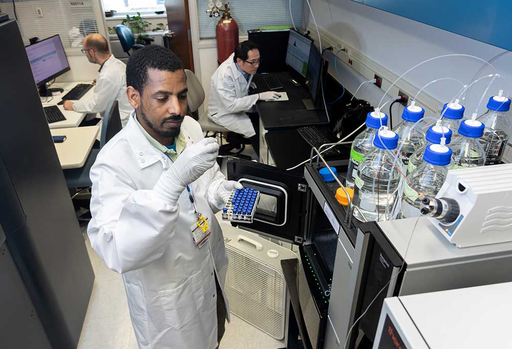
Transforming discovery to care.
Our researchers are driven by their limitless curiosity to discover new and better ways to make these children better today and healthier tomorrow.

We're focused on improving child health through exceptional patient care, groundbreaking research, continuing education, and outreach and prevention.

Then we're looking for you! Work at a place where you can change lives...including your own.

When you give to Arkansas Children's, you help deliver on our promise of a better today and a healthier tomorrow for the children of Arkansas and beyond
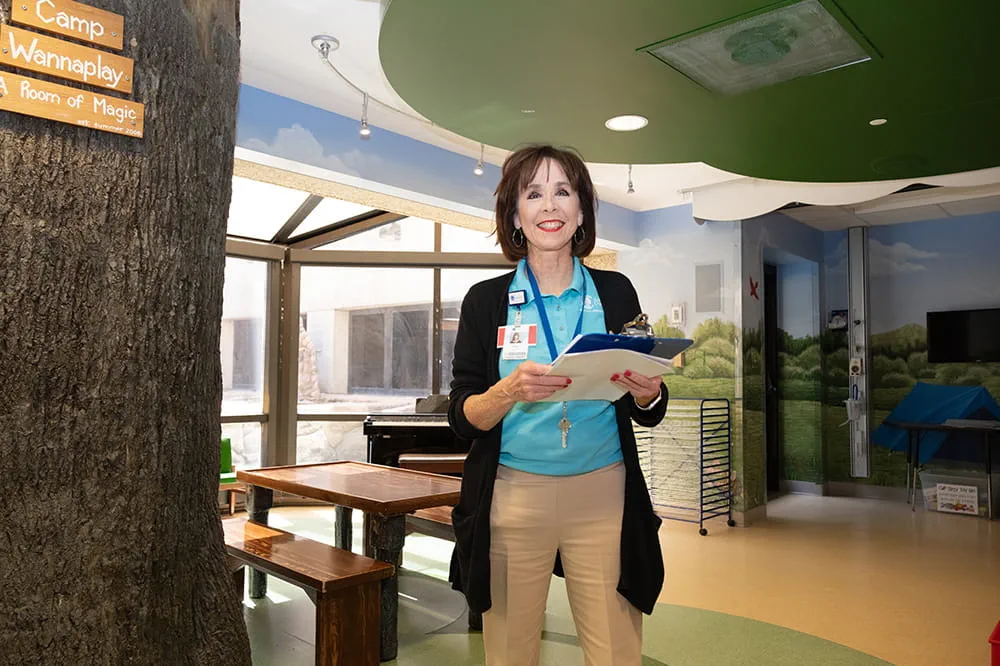
Become a volunteer at Arkansas Children's.
The gift of time is one of the most precious gifts you can give. You can make a difference in the life of a sick child.

Join our Grassroots Organization
Support and participate in this advocacy effort on behalf of Arkansas’ youth and our organization.

Learn How We Transform Discovery to Care
Scientific discoveries lead us to new and better ways to care for children.

Learn How We Transform Discovery to Care
Scientific discoveries lead us to new and better ways to care for children.

Learn How We Transform Discovery to Care
Scientific discoveries lead us to new and better ways to care for children.

Learn How We Transform Discovery to Care
Scientific discoveries lead us to new and better ways to care for children.

Learn How We Transform Discovery to Care
Scientific discoveries lead us to new and better ways to care for children.

Learn How We Transform Discovery to Care
Scientific discoveries lead us to new and better ways to care for children.

When you give to Arkansas Children’s, you help deliver on our promise of a better today and a healthier tomorrow for the children of Arkansas and beyond.
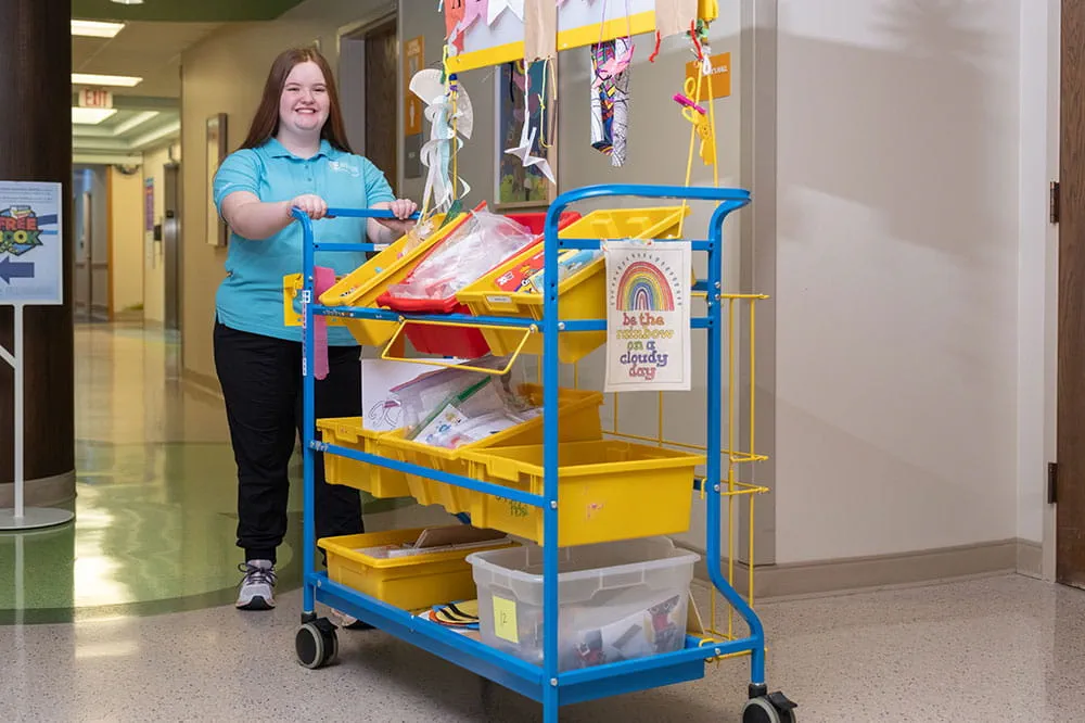
Your volunteer efforts are very important to Arkansas Children's. Consider additional ways to help our patients and families.

Join one of our volunteer groups.
There are many ways to get involved to champion children statewide.

Make a positive impact on children through philanthropy.
The generosity of our supporters allows Arkansas Children's to deliver on our promise of making children better today and a healthier tomorrow.

Read and watch heart-warming, inspirational stories from the patients of Arkansas Children’s.

Hello.

Arkansas Children's Hospital
General Information 501-364-1100
Arkansas Children's Northwest
General Information 479-725-6800

Ventricular Septal Defect (VSD)
What is a ventricular septal defect?
A ventricular septal defect (VSD) is a hole between the two lower chambers of the heart, called the ventricles. It is a type of congenital heart condition, which means it’s a condition a baby is born with. VSDs are one of the most common congenital heart conditions.
The size of a VSD can vary from small (<3mm in diameter) to large (>6 - 10mm in diameter); there may also be more than one. A small VSD may not cause any problems and may even close on its own. Small VSDs account for 80-85% of all VSDs and close spontaneously. Larger VSDs cause more blood to flow through the lungs than normal. Over time, this can cause the following symptoms:
- failure to thrive (poor growth)
- heart and respiratory failure, requiring an intervention within 3-6 months of life
- heart and lung stress
- heart failure
- pulmonary hypertension (high blood pressure in the lungs)
There are five basic types of ventricular septum classifications:
- Perimembranous (Paramembranous, Membranous): VSD immediately below and adjacent to the commissure between the anterior and septal leaflets of the tricuspid valve. It is closely associated with the bundle of His (conduction system of the heart) at its posterior and inferior corners. This defect often lies under the commissure between the non- and right-leaflets of the aortic valve can cause aortic valve damage.
- Conoventricular (Subaortic, Infracristal): VSD frequently associated with malalignment of the conal septum (i.e., Tetralogy of Fallot with anterior malalignment, interrupted aortic arch with posterior malalignment). This defect extends more superiorly and anteriorly than a perimembranous VSD, but is also closely associated with the bundle of His (conduction system of the heart).
- Subpulmonary (Supracristal, Conal, Intraconal): Defect in the conal septum, which lies immediately below the pulmonary valve and adjacent to the right coronary cusp of the aortic valve. This VSD has a high risk of damaging the aortic valve and requires surgical repair.
- Inlet (Atrioventricular Canal Type): VSD immediately below the atrioventricular valves (septal leaflet of the tricuspid/right AV valve). Commonly associated with a primum ASD and also may be related to a separate perimembranous VSD.
- Muscular: VSD occurring in the muscular septum between the two ventricles of the heart. Many of these defects either close over time or are amenable to device closure.
What are the signs and symptoms of a ventricular septal defect?
The symptoms of a VSD can vary depending on its size and where it is located. Many children with a VSD don’t have any symptoms, especially if it is small. If your child does have symptoms they may include:
- Heart murmur (a sound heard by your baby’s doctor with a stethoscope)
- Poor growth
- Fast breathing
What causes a ventricular septal defect?
VSDs develop early in pregnancy when the heart is formed. It is not known what causes VSDs to develop.
How are ventricular septal defects treated?
The surgical approach involves a standard midline incision (sternotomy). A machine that allows the heart and lungs to rest (cardiopulmonary bypass) allows the surgery team to repair the area of concern. Surgical repair most commonly involves placing a patch of material, either biological or synthetic, to close the ventricular septal defect.
How is a ventricular septal defect treated?
Your child’s treatment will depend on the size of the VSD and where it is located. Children with small VSDs may not need any treatment. Instead, your child’s doctor may track their condition over time. Many small VSDs will close on their own. Your care team at Arkansas Children’s is experienced in treating ventricular septal defects and will work with you to develop the best treatment plan for your child’s specific condition and symptoms. Treatments may include:
- Medicines to reduce fluid in the lungs.
- A high-calorie formula or feeding tube to help your child get enough nutrition to grow.
- Cardiac catheterization using a long, small tube (catheter) inserted into a vein and guided to the heart to place a special device over the VSD to close it.
- Surgery to close the VSD using stiches or a special patch.
The average length of hospital stay is around 5-7 days.

Appointments
New and existing patients can visit our appointment hub for several ways to request an appointment, including online scheduling for many services.
Request an appointment
